Examinations
EXAMINATIONS
We offer the communities of Narrabri, Coonabarabran, Quirindi and Wee Waa a full range of ultrasound examinations. Using state-of-the-art equipment we provide:
- General ultrasound – abdominal, renal, pelvic
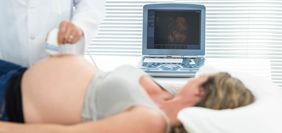
- Obstetric ultrasound – dating, nuchal translucency (12 week scan), morphology scan (19-20 week scan), third trimester scan (fetal well being)
- Vascular ultrasound – arm and leg deep vein thrombosis (DVT), Doppler scans (leg, carotid and renal arteries)
- Musculoskeletal ultrasound – all types
- Small parts ultrasound – thyroid, scrotum, neck, face, salivary glands
What is ultrasound?
An ultrasound scan uses high-frequency sound waves to make an image of a person’s internal body structures. Doctors commonly use ultrasound to study a developing unborn baby, a person’s abdominal and pelvic organs, muscles and tendons, or their heart and blood vessels.
Patients are not exposed to ionizing radiation making ultrasound safer than diagnostic techniques such as X-rays and CT scans. In fact, there are no known harmful effects when used as directed by your health care provider.
The ultrasound machine directs high-frequency sound waves at the internal body structures being examined. The reflected sounds, or echoes, are recorded to create an image that can be seen on a monitor. The sound waves are emitted and received from a small, hand-held probe. The high frequency of the sound means the human ear cannot hear it – which is why it is called ultrasound.
An ultrasound scan is usually non-invasive (done from outside the body). However, some scans are done with a special probe that is inserted into the patient’s vagina (for some obstetric or pelvic examinations). Sometimes, doctors will work with sonographers to do low risk procedures such as injecting hips, knees, shoulders, elbows or wrists to relieve pain.
What does the ultrasound procedure involve?
If you are having an upper abdomen ultrasound, you will need to lie down on an examination table or bed. The sonographer will place some gel onto your skin to provide better contact between your body and the ultrasound probe. They will then place the hand-held probe on your skin above the area of your body, organ or tissue that is being studied.
The two-dimensional (or sometimes three-dimensional) pictures are shown instantly on a monitor.
Other types of ultrasound may need a slightly different procedure. For example, a woman undergoing an investigation of her pelvis may have a transvaginal scan, which involves inserting a special ultrasound probe into the vagina rather than (or as well as) scanning through the front of the pelvis.
The average ultrasound scan takes around 20 minutes, depending on the type of examination. After the procedure, the sonographer will give you paper towels to wipe off the gel, and you can then get dressed. The results of the ultrasound scan are sent to your doctor.